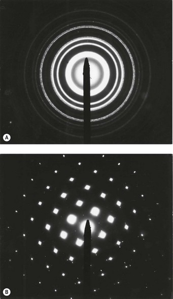

Part of a crystal structure, and its simulated diffraction pattern. By combining a simulated pattern with an observed diffraction image, you can auto-index the pattern and determine the orientation of your crystal. Through the analysis of TEM data, we can get the information of the sample size, particle size statistics, crystal grain size, crystal structure, crystal phase. Simulating electron diffraction for scapolite along 111. SingleCrystal lets you simulate X-ray, neutron and electron diffraction patterns from single crystals, display reciprocal lattice sections and construct stereographic projections of planes or vectors.
#Crystalmaker electron diffraction pattern example series#
The length and width of the electron transparent region of the lamella are approximately 5 m and 3 m, respectively. TEM data analysis refers to a data processing method to obtain sample structure information through a series of measurements, analysis, and calibration of the original spectrum. 4.4 EDS spectrum of a straight nanowire from sample 2 with labeled peaks.

c A TEM overview of the lamella (show in Fig. yield during growth and preliminary transmission electron microscopy (TEM). Thereby, not only the d-spacings but also the positions of the diffraction. A metastable hexagonal ω-Fe phase with its particle size of about 1-2 nm coexists at the m1 \)) structures depending upon the extent of collapse of the atomic planes. a, b Show the schmatic of imaging and diffraction patterns. Electron diffraction pattern from the area marked in c.


 0 kommentar(er)
